"The Johns Hopkins Atlas of Human Functional Anatomy" by Leon Schlossberg and George D. Zuidema is a comprehensive resource focused on **human anatomy and physiology** 【4】【6】.
Key themes and topics covered in the book include:
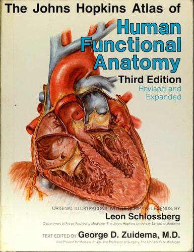
* **Detailed anatomical illustrations**: The atlas features numerous color illustrations, with over 200,000 copies sold, depicting all organs and systems of the human body 【3】【5】. These illustrations are created by medical artist Leon Schlossberg 【1】【3】.
* **Functional anatomy**: The book correlates each body structure with its functions within different systems and relates it to the human body as a whole 【1】.
* **Expert-written descriptions**: The text accompanying the illustrations is written by current and former faculty from the Johns Hopkins University School of Medicine, with each section on a particular part of the anatomy authored by an expert in that field 【2】【3】.
* **Systemic and organ-specific coverage**: The atlas covers all systems and organs of the human body 【1】. Specific enhancements in the fourth edition include new chapters and plates on the aorta, liver, thymus, breast, prostate, and hernias 【2】【7】. It also features a detailed illustration of the interior of the male anatomy 【2】【8】.
* **Anatomical, physiological, and clinical terms**: The atlas includes an extensive glossary of these terms 【1】.
* **Indices for quick reference**: An index of plates and descriptions is provided for easy identification of any structure, organ, or system 【2】【3】.
The book is considered a trusted and authoritative source for general readers and students at all levels 【3】【5】. It has been published in multiple editions, with topics including human anatomy, human anatomy atlases, anatomy atlases, and physiology atlases 【4】【6】.